Types of Pregnancy Ultrasounds: A Week-by-Week Guide for Expecting Parents
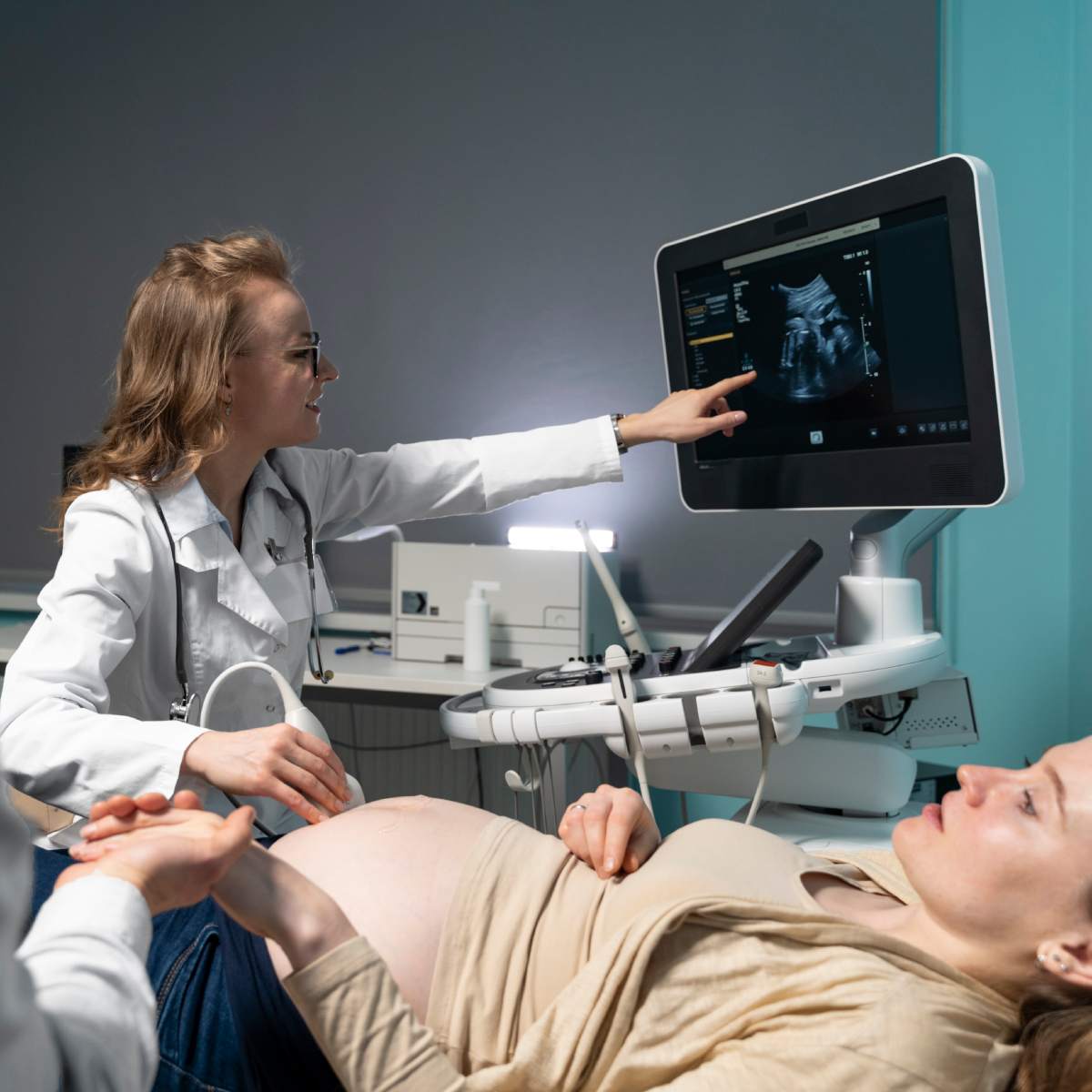
Table of Contents
Over 95% of pregnant women worldwide will undergo at least one ultrasound scan during pregnancy, making it one of the most common and safest diagnostic tools used in modern prenatal care.
Regular ultrasound scans during pregnancy are an important part of tracking the health and development of your unborn child. These non-invasive examinations use sound waves to produce images of your baby in the womb. But did you know that there are various types of ultrasound scans during pregnancy? These tests are performed based on the stage of the pregnancy and for a variety of reasons, such as determining the chance of preterm labor and Down syndrome, and many others.
Being aware of the numerous types of prenatal ultrasounds and when they are advised can make you feel better prepared and more informed during your pregnancy.
What is an Ultrasound Scan?
An ultrasound is a highly safe method that employs sound waves to make images of your baby when you are pregnant. There is no use of radiation or needles. According to scientific studies, ultrasound scans are considered safe for mother and baby when performed by qualified professionals, and there is no evidence that routine ultrasound exposure causes harm. However, unnecessary or non-medical ultrasounds (such as “keepsake” scans) should be avoided.
How Does Ultrasound Work?
When an ultrasound is performed during pregnancy, the sonographer, who performs the ultrasound, applies gel to your abdomen. Your skin is covered with a transducer, a portable wand that is softly moved around. Sound wave pulses travel from the transducer to the uterus, or womb, where your unborn child is present. The ultrasound equipment receives the echoes produced by the sound waves. These soundwave echoes are then transformed into images by a computer.
Ultrasound images can be viewed in real time, allowing the operator to observe the movement, heartbeat, and anatomy of the fetus and surrounding organs. This technology can also be used in Doppler mode to measure blood flow through the umbilical cord or fetal vessels, which is useful for assessing fetal well-being.
Types of Ultrasound Scans and Their Recommended Timing
Below is a comprehensive table of ultrasound types, their timing, and their main purpose:
| Ultrasound Type | Typical Timing | Description/Purpose |
| Transvaginal Ultrasound | 5–8 weeks (and as needed) | Used in very early pregnancy for dating, viability, and to check for ectopic pregnancy. The probe is inserted into the vagina for clearer images of early development. |
| Dating Scan (Viability Scan) | 7–12 weeks | Estimates gestational age, confirms intrauterine pregnancy, and detects number of embryos. |
| Nuchal Translucency (NT) Scan | 11–14 weeks | Measures fluid at the back of baby’s neck for Down syndrome risk; part of first trimester screening. |
| Anatomy Scan (Morphology/Level II) | 18–22 weeks | Assesses fetal anatomy, growth, organ development, detects most major birth defects, measures placenta and amniotic fluid. |
| Fetal Echocardiography | 18–24 weeks (if indicated) | Detailed examination of fetal heart structure and function, often recommended if there is a family history of heart defects or abnormal findings in the anatomy scan. |
| Growth Scan (Wellbeing Scan) | 28–32 weeks (or as needed) | Assesses fetal growth, amniotic fluid, placental function, fetal movements, and position. |
| Doppler Ultrasound | Any time after 24 weeks (as indicated) | Measures blood flow in the umbilical cord, placenta, and fetal vessels, important in high-risk pregnancies, suspected growth restriction, or preeclampsia. |
| Biophysical Profile (BPP) | After 32 weeks (as needed) | Combines ultrasound and non-stress test (NST) to check baby’s movements, breathing, tone, amniotic fluid, and heart rate. Used in late pregnancy or if complications arise. |
| 3D/4D Ultrasound | 24–34 weeks (elective/if indicated) | Provides three-dimensional images and real-time motion; useful for certain facial or limb anomalies, but not part of routine care. |
| Presentation/Position Scan | 36–40 weeks | Confirms baby’s position (head down, breech) and placental location before delivery. |
Other specialized ultrasounds, such as targeted scans for specific fetal abnormalities, uterine artery Doppler, or cervical length measurement, may be recommended based on your pregnancy’s unique needs or risk factors.
Why Should I Have an Ultrasound Scan?
An ultrasound scan can give you and your medical team more details about your pregnancy and fetus. Your further care will be guided by the results of the study. The following can be checked with an ultrasound, depending on the type and number of weeks you are pregnant:
- The heartbeat and overall health of your unborn child
- Whether you are expecting multiples, triplets, or twins
- Your due date
- Your baby’s position in the womb
- Your cervix
- Your placenta's location
- The size and growth of the fetus
Ultrasound scans are also essential for guiding certain prenatal procedures, such as amniocentesis or chorionic villus sampling (CVS), and for monitoring high-risk pregnancies.
Questions to Ask Your Doctor
While having an ultrasound scan during pregnancy, there are many questions that may come up in your mind. You can ask your doctor or midwife some of these questions:
- Why would you recommend this ultrasound?
- What are the advantages of this ultrasound procedure?
- If I decide not to get the ultrasound, are there any risks?
- What is involved in the ultrasound?
- Can an ultrasound scan harm my baby?
- When can I expect to receive the results?
Bottom Line
Ultrasound scans can give you and your midwife or doctor information about the status of your pregnancy and the health of your unborn child. These scans may tell you that your baby is developing normally, but they may potentially reveal an abnormality. You can discuss additional testing with your doctor or midwife if the ultrasound results raise any health issues.
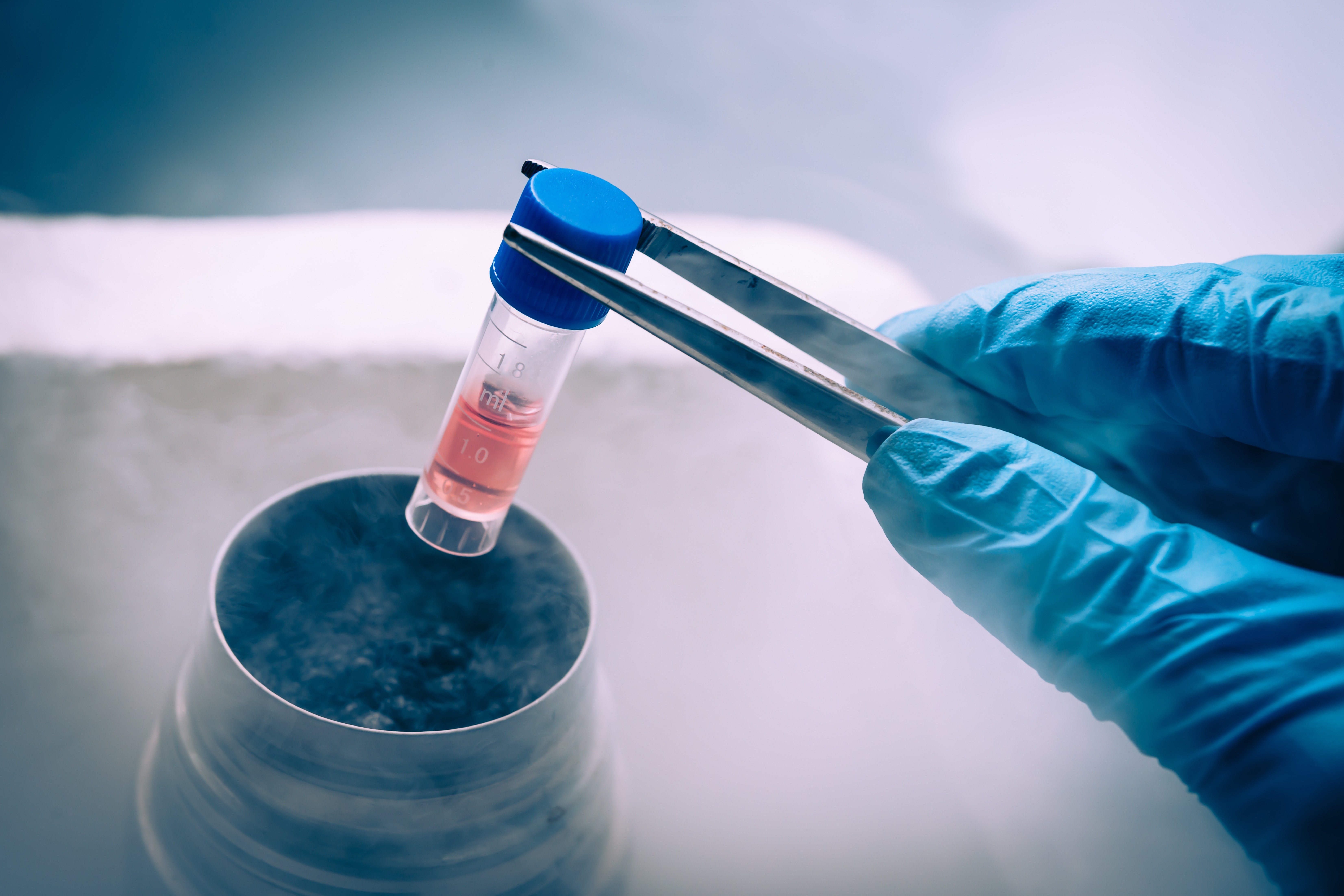
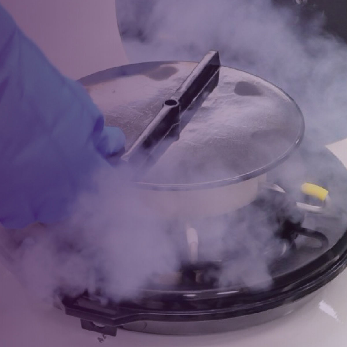
Also, if you’re looking to save your baby’s and your family’s health, storing your baby’s cord blood is an excellent decision. By saving the precious stem cells present in the cord blood, you secure the future health of your unborn baby. Sign in today with Cryoviva Life Sciences, and gain multiple benefits of treating life threatening diseases.
.jpg)
.jpg)


.jpg)


 Enquiry
 Email
Email Phone
Phone
 Whatsapp